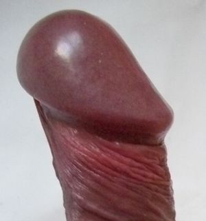
Glans penis
In male human anatomy, the glans penis (or simply glans, /ɡlænz/) is the sensitive bulbous structure at the distal end of the penis.[1] The glans is anatomically homologous to the clitoral glans of the human female. Typically, the glans of the normal penis is completely or partially covered by the foreskin, though the foreskin can generally be retracted over and past the glans.

The glans penis, or simply the glans, refers to the head of the penis. Nature treated the glans penis as an internal organ, so the epithelium is mucosa and provided the foreskin for protection against infection and trauma. The glans is normally covered by a hood of folded skin called the foreskin. The foreskin moisturizes the glans penis by transudation. The surface of the glans is mucosal tissue that, under normal conditions in the preputial sac, is moist and supple.
The glans penis receives much of its blood supply through the frenular artery. If the frenular artery is damaged by circumcision, the glans penis of some men may not get full erection.
Nature has designed the glans penis to be an internal organ, therefore the foreskin should be kept in position to cover the glans except for sexual activity and occasional washing.
First retraction of the foreskin
Functions of the foreskin include protecting the glans penis from abrasion, keratinization and moistening it by with body oils and moisture to keep it moist, smooth, and glossy with full sensation.[2] The glans of the foreskinned penis under the foreskin normally is kept moist to wet with a mixture of moisture and skin oil by transudation. The foreskin will also collect pre-ejaculate when it is secreted. When the foreskin is first retracted, There may be a substantial accumulation of beneficial smegma that has been collecting there harmlessly since birth.
It is normal for the glans penis (head of the penis) to be very sensitive to touch when the foreskin is retracted for the first time since birth.
There is no particular reason to touch the glans so, if it is painful to touch, then one may simply avoid touching it. Nature provides the foreskin with its well known protective functions. Simply keeping the foreskin forward in its protective position over the glans penis will prevent pain.
Sensitivity and innervation of the glans penis
Cepeda-Emiliani et al. (2023) reported the glans penis has dual innervation from both the dorsal and perineal nerves.[3]
Cepeda-Emiliani et al. (2025) reported on the sensory corpuscles found in the glans and frenular region:
A wide variety of sensory corpuscles in the glans and frenular region were spatially segregated in a highly organized manner (Figures 8–10 and Figure S4). These included genital, Krause, Pacinian, Paciniform, Golgi-Mazzoni, Ruffini-like, Meissner, and numerous other corpuscles that were difficult to classify.[4]
When the glans penis loses the protection of the foreskin it collects a layer of keratin and loses its glossy appearance. The thick layer of keratin drastically reduces sensitivity.
The dartos muscle sheet in the foreskin produces contractions that are slow, sustained, and may produce great force, such as in cold temperatures.[5]
The innervation of the glans responds to pressure, so the glans is meant to be stimulated with the foreskin as its medium.
While the human glans penis is protopathic, the prepuce contains a high concentration of touch receptors in the ridged band.
In the human penis, the prepuce is known to have ten times more corpuscular sensory receptors than the glans penis.
The glans is insensitive to light touch, heat, cold, and even pinpricks, as researches at the Department of Pathology in the Health Science Centre at the University of Manitoba discovered.[6] The corona of the glans contains scattered free nerve endings, genital end bulbs, and pacinian corpuscles, which transmit sensations of pain and deep pressure. The glans is nearly incapable of detecting light touch.
An impressive study performed by objective Chinese researchers conclusively demonstrated that circumcision reduces the glans sensitivity to vibration. "The test group were 1.97 +/- 0.71, 2.64 +/- 1.38, 3.09 +/-1.46 and 2.97 +/- 1.20 respectively before and 1, 2 and 3 months after circumcision, with significant difference between pre- and post-operation (P < 0.05).[7]
The protection provided by the foreskin prevents keratinization and helps to maintain sensitivity of the glans penis. Foreskinned men frequently report that they feel pain when their glans penis is touched with their fingers, so many foreskinned men prefer to keep their glans penis covered by their intact foreskin except for occasional washing and sexual activity.
The glans penis after circumcision of the protective foreskin
If left uncovered for long periods of time, the surface of the glans becomes desiccated. In the circumcised penis, the glans is permanently exposed; the surface of the glans dries out and develops layers of keratin and loses sensation. The experience of many men indicates that the heavy layer of keratin on the glans penis develops a cracked appearance and feels hard when touched as compared with the normal glans pens.
The glans penis after restoration of the protective foreskin
There is a paucity of scientific study of the restored penis, so this information is based on the experience of many restored men.
Some men have reported that, after the glans penis is covered by a restored foreskin, they experienced peeling of a layer of keratin similar to the peeling that occurs after sunburn, however that is only the beginning of restoration of the glans penis.
As foreskin restoration proceeds and more foreskin is available, the restored foreskin will start to cover the glans penis and the glans penis usually is bathed with natural skin oil and moisture by transudation in a manner similar to an intact penis. Over a period of several years, dekeratinization will continue and the glans will become softer, glossier, and more sensitive. There usually will be a return to more normal color. One cannot say how long this process will take due to a lack of data.
Excessive washing of the restored preputial sac removes oil and should be avoided.[8]
See also
External links
 Halata Z, Munger BL. The Neuroanatomical Basis for the Protopathic Sensibility of the Human Glans Penis. Brain Research. 23 April 1986; 371(2): 205-30. PMID. DOI. Retrieved 23 October 2025.
References
- ↑

glans penis
, The Free Dictionary by Farlex. Retrieved 25 January 2025. - ↑
 Fleiss P, Hodges F, Van Howe RS. Immunological functions of the human prepuce. Sex Trans Infect. October 1998; 74(5): 364-67. PMID. PMC. DOI. Retrieved 14 January 2022.
- ↑
 Cepeda-Emiliani A, Gándara-Cortés M, Otero-Alén M, García H, Suárez-Quintanilla J, García-Caballero T, Gallego R, García-Caballero R. Immunohistological study of the density and distribution of human penile neural tissue: gradient hypothesis. Int J Impot Res. 2 May 2023; 35(3): 286-305. PMID. DOI. Retrieved 24 November 2023.
- ↑
 Cepeda-Emiliani A, Otero-Alén M, Suárez-Quintanilla J, Gándara-Cortés M, García-Caballero T, García-Caballero R, García-Caballero L. The sensory penis: A comprehensive immunohistological and ontogenetic exploration of human penile innervation. Andrology. 19 September 2025; : 1-41. PMID. DOI. Retrieved 1 October 2025.
- ↑ Jefferson G. The peripenic muscle; some observations on the anatomy of phimosis. Surgery, Gynecology and Obstetrics 1916 Aug;23(2):177-81.
- ↑
 Taylor JR, Lockwood AP, Taylor AJ. The prepuce: specialized mucosa of the penis and its loss to circumcision. Br J Urol. 1996; 77: 291-5. PMID. DOI. Retrieved 23 September 2019.
- ↑
 Yang D, Lin H, Zhang B, Guo W. [Circumcision affects glans penis vibration perception threshold]. Zhonghua Nan Ke Xue. April 2008; 14(4): 328-30. PMID. Retrieved 14 March 2024.
Quote:https://www.ncbi.nlm.nih.gov/pubmed/18481425?dopt=Abstract
- ↑
 Birley HDL, Walker MM, Luzzi GA, Bell R, et al. Clinical Features and management of recurrent balanitis; association with atopy and genital washing
  . Genitourin Med. October 1993; 69(5): 400-3. PMID. PMC. DOI. Retrieved 25 January 2025.
